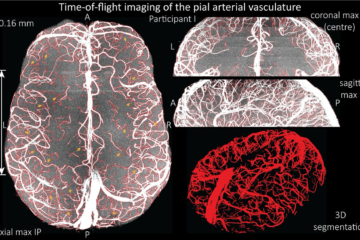
For this year’s ISMRM, I’ve investigated how we can image the pial arterial vasculature using a time-of flight contrast. Surprisingly, we found that we are not limited by physiological parameters, but ‘simply’ by the voxel size. All the gory details can be found in the video below. Note that this is a slightly longer version than the original ISMRM presentation with a bit more information.
All the acquisitions were performed on Siemens 7T scanners. To reproduce the 0.16 mm acquisition, I have collected all the important parameters below. I have used the product TOF implementation, but had to increase the maximum limit of the base resolution (default is 1024) in the sequence code. The TONE ramp is set to 100%, which – according to my understanding – simply turns it off, because we don’t need it for pial arteries.
- acquisition time: 11 min 16 seconds
- resolution: 0.16 mm x 0.16 mm x 0.16 mm
- orientation: transversal
- phase encoding direction: R >> L
- phase oversampling: 0%
- slice oversampling: 15.4%
- slices per slab: 52
- FOV read: 204 mm
- FOV phase: 85.2%
- slice thickness: 0.16 mm
- TR: 20 ms
- TE 6.56 ms
- flip angle: 18 degree
- base resolution: 1280
- PAT mode: GRAPPA
- acceleration factor PE: 2
- reference lines PE: 32
- POCS: read & slice
- TONE ramp: 100%
- 3D centric reordering: on
- dimension: 3D
- asymmetric echo: allowed
- bandwidth: 100Hz/Px
- flow compensation: yes
Please let me know how you go, if you can reproduce the results or run into any problems. Also, if any parameter is missing, please get in touch!
0 Comments